NCLEX Cardiac Nursing Essentials: Heart Conditions, Meds & Priority Interventions
Cardiac nursing is one of the highest-yield content areas on the NCLEX, spanning heart failure, acute coronary syndrome, arrhythmias, valvular disorders, and the medications used to manage them. If you understand how the heart works, how it fails, and what the nurse does about it, you will be prepared for a significant portion of your exam questions. This guide covers the NCLEX cardiac nursing essentials you need to master — from assessment and lab values to priority interventions and delegation.
Think hemodynamics, not just heart disease
The NCLEX tests your ability to connect cardiac pathophysiology to nursing action. Every cardiac condition ultimately affects cardiac output — the volume of blood the heart pumps per minute. When cardiac output drops, tissue perfusion suffers. Your job as the nurse is to recognize the signs early, intervene to restore perfusion, and prevent life-threatening complications.
Heart Failure: Left-Sided vs. Right-Sided
Heart failure is arguably the most commonly tested cardiac topic on the NCLEX. The key to mastering heart failure questions is understanding which side of the heart is failing and where the blood backs up as a result.
Left-sided heart failure causes blood to back up into the pulmonary vasculature. The hallmark signs are all respiratory: dyspnea, orthopnea, paroxysmal nocturnal dyspnea, crackles (rales) on auscultation, pink frothy sputum (indicating pulmonary edema), and tachycardia. The patient may need to sleep propped up on multiple pillows or in a tripod position. Nursing priorities include elevating the head of the bed to at least 30–45 degrees, administering prescribed diuretics (furosemide is the go-to), monitoring oxygen saturation, auscultating lung sounds every shift, and tracking strict intake and output with daily weights.
Right-sided heart failure causes blood to back up into the systemic venous circulation. Look for jugular vein distention (JVD), peripheral edema (especially in the ankles and feet), hepatomegaly, ascites, and weight gain from fluid retention. Right-sided failure most commonly results from chronic left-sided failure, but it can also be caused by pulmonary hypertension or COPD (cor pulmonale). Nursing priorities include monitoring daily weights (a gain of more than 2 pounds in 24 hours or 5 pounds in a week suggests fluid retention), sodium restriction (typically less than 2 grams per day), and monitoring potassium levels carefully when the patient is on diuretics.
A critical NCLEX tip: when a question describes a patient with both crackles in the lungs AND peripheral edema, the patient has biventricular failure (both sides). The priority intervention is always the life-threatening issue first — address the pulmonary edema (left-sided symptoms) before managing the peripheral edema.
Acute Coronary Syndrome: Angina and Myocardial Infarction
Acute coronary syndrome (ACS) is an umbrella term that includes unstable angina, NSTEMI (non-ST-elevation MI), and STEMI (ST-elevation MI). All three involve reduced blood flow to the myocardium, but they differ in severity and the degree of cardiac tissue damage.
The classic presentation of ACS is substernal chest pain — often described as pressure, squeezing, or heaviness — that may radiate to the left arm, jaw, neck, or back. The patient is typically diaphoretic, nauseated, and short of breath. However, the NCLEX also tests atypical presentations: women, diabetic patients, and the elderly may present with fatigue, epigastric discomfort, jaw pain, or dyspnea only, without classic chest pain.
For initial nursing interventions during chest pain, remember the MONA framework (though current evidence-based practice has updated the order of priority):
- Aspirin — 160-325 mg, chewed immediately (antiplatelet effect is the top priority)
- Nitroglycerin — sublingual, up to 3 doses at 5-minute intervals; hold if systolic BP is below 90 mmHg
- Oxygen — only if SpO2 is below 90%, or if the patient has respiratory distress, heart failure, or shock. Do not give routine oxygen to a patient with normal oxygen saturation — per 2025 ACC/AHA ACS guidelines, routine supplemental O2 is not recommended in normoxic patients
- Morphine — for pain unrelieved by nitroglycerin (used cautiously due to hypotension risk)
Troponin is the most specific cardiac biomarker. Elevated troponin I or troponin T indicates myocardial injury and supports MI diagnosis when paired with symptoms, ECG changes, and clinical findings — it differentiates MI from unstable angina. Serial troponin levels are drawn at presentation, 3 hours, and 6 hours. CK-MB rises and falls more rapidly and helps identify reinfarction if troponin is already elevated.
Cardiac Arrhythmias: What the NCLEX Expects You to Know
The NCLEX does not expect you to be a cardiologist, but you must be able to recognize life-threatening arrhythmias and know the immediate nursing response. Focus on these key rhythms:
Sinus bradycardia — heart rate below 60 bpm with a normal rhythm pattern. Asymptomatic bradycardia needs no treatment; symptomatic bradycardia (hypotension, dizziness, syncope) is treated with atropine. If atropine is ineffective, transcutaneous pacing may be required.
Atrial fibrillation (A-fib) — an irregularly irregular rhythm with no identifiable P waves. The biggest risk is thrombus formation in the fibrillating atria, leading to stroke. Patients on chronic A-fib are placed on anticoagulants (warfarin or DOACs like apixaban and rivarelbaban). For rate control, beta-blockers (metoprolol) or calcium channel blockers (diltiazem) are first-line. Monitor INR for patients on warfarin — therapeutic range is 2.0–3.0.
Ventricular tachycardia (V-tach) — if the patient has a pulse, administer amiodarone and prepare for synchronized cardioversion. If the patient is pulseless, this is a cardiac arrest — begin CPR and prepare for defibrillation.
Ventricular fibrillation (V-fib) — always pulseless, always an emergency. The priority is immediate defibrillation. Begin high-quality CPR while the defibrillator is being prepared. Administer epinephrine every 3–5 minutes during the code.
Asystole — flatline, no electrical activity. Asystole is NOT shockable. The intervention is CPR and epinephrine. Do not defibrillate asystole — this is a common NCLEX distractor.

Shockable vs. non-shockable rhythms
The NCLEX loves to test whether you know which rhythms are shockable. Ventricular fibrillation and pulseless ventricular tachycardia are shockable — use a defibrillator. Asystole and pulseless electrical activity (PEA) are non-shockable — the intervention is CPR and epinephrine. Knowing this distinction can earn you multiple exam points.
Essential Cardiac Medications for the NCLEX
Cardiac pharmacology is heavily tested. You need to know the drug class, primary action, key nursing considerations, and what to monitor for each of these medication groups:
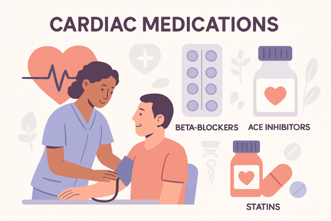
ACE Inhibitors (lisinopril, enalapril — endings in "-pril") — reduce afterload by preventing the conversion of angiotensin I to angiotensin II. First-line for heart failure and hypertension. Monitor for hyperkalemia, dry persistent cough (a hallmark side effect), and angioedema. ACE inhibitors increase potassium — use caution with potassium supplements, salt substitutes, and potassium-sparing diuretics such as spironolactone. If combined (e.g., in selected HFrEF patients), potassium and renal function must be closely monitored; MRAs are generally appropriate only when eGFR is >30 and potassium is <5.0 mEq/L. Check renal function (BUN/creatinine) regularly. Question the dose or notify the provider if potassium is ≥5.0 mEq/L — follow provider/facility hold parameters.
Beta-Blockers (metoprolol, atenolol, carvedilol — endings in "-olol") — reduce heart rate, decrease myocardial oxygen demand, and lower blood pressure. Used in heart failure, post-MI, and arrhythmias. Monitor for bradycardia and hypotension. Teach patients never to stop beta-blockers abruptly — taper gradually to prevent rebound tachycardia. Hold if heart rate is below 60 bpm or systolic BP is below 100 mmHg.
Digoxin — increases the force of cardiac contraction (positive inotrope) and slows the heart rate. Used in heart failure and atrial fibrillation. The therapeutic range is narrow: 0.5–2.0 ng/mL. Check the apical pulse for one full minute before administration — hold and notify the provider if the rate is below 60 bpm. Signs of digoxin toxicity include nausea, vomiting, visual disturbances (yellow-green halos), and bradycardia. Hypokalemia increases the risk of toxicity, so always check potassium before giving digoxin.
Anticoagulants — warfarin (oral, monitored by INR, therapeutic range 2.0–3.0, antidote is vitamin K) and heparin (IV or subcutaneous, monitored by aPTT, therapeutic range 1.5–2.5 times control, antidote is protamine sulfate). Direct oral anticoagulants (DOACs) such as apixaban and rivaroxaban do not require routine lab monitoring but have limited reversal options.
Nitroglycerin — causes vasodilation, which decreases preload and relieves chest pain. Administer sublingually; the patient should feel a tingling sensation under the tongue. Store in a dark glass container. Do not administer if the patient has taken a PDE5 inhibitor (sildenafil/tadalafil) within the past 24–48 hours due to the risk of severe hypotension.
Know your cardiac medication hold parameters
The NCLEX frequently tests when to hold a medication. Hold beta-blockers if HR is below 60 or BP is below 100/60. Hold digoxin if apical pulse is below 60. For ACE inhibitors, question the dose or notify the provider if potassium is ≥5.0 mEq/L — follow provider/facility hold parameters. Hold nitroglycerin if systolic BP is below 90. These hold parameters appear repeatedly on the exam.
Cardiac Lab Values and Diagnostics
You must know these lab values for cardiac NCLEX questions:
- Troponin I/T — most specific marker for myocardial injury. Normal is less than 0.04 ng/mL. Elevated troponin indicates myocardial injury and supports MI diagnosis when combined with symptoms, ECG changes, and clinical context. Note: troponin can also rise with myocarditis, renal disease, heart failure, sepsis, and pulmonary embolism.
- BNP (B-type natriuretic peptide) — released by the ventricles when they are stretched by volume overload. BNP >100 pg/mL may suggest heart failure, but diagnosis requires clinical assessment and other tests. BNP can also be affected by age, kidney disease, and obesity. Higher BNP levels generally correlate with more severe volume overload, but clinical context is essential for interpretation.
- INR — monitors warfarin therapy. Therapeutic range is 2.0–3.0 (2.5–3.5 for mechanical heart valves).
- aPTT — monitors heparin therapy. Therapeutic range is 1.5–2.5 times the control value (typically 60–80 seconds).
- Potassium — normal 3.5–5.0 mEq/L. Hypokalemia and hyperkalemia both cause cardiac arrhythmias. Always check potassium before giving digoxin or potassium-wasting diuretics.
- Magnesium — normal 1.5–2.5 mEq/L. Low magnesium contributes to arrhythmias (especially torsades de pointes) and makes hypokalemia resistant to correction.
For diagnostic tests, know that a 12-lead ECG is the first-line diagnostic for chest pain and arrhythmias. An echocardiogram assesses ventricular function and ejection fraction (normal is 55–70%). Cardiac catheterization is the gold standard for diagnosing coronary artery disease and may be followed by percutaneous coronary intervention (PCI) with stent placement.
Cardiac Assessment: What to Prioritize
A thorough cardiac assessment on the NCLEX follows the ABCs and focuses on tissue perfusion. Key assessment findings to monitor include:
- Vital signs — heart rate, blood pressure, respiratory rate, oxygen saturation. Note trends, not just single readings.
- Heart sounds — S1 and S2 are normal. An S3 sound (ventricular gallop) is associated with heart failure. An S4 sound (atrial gallop) is associated with stiff, hypertrophic ventricles.
- Peripheral perfusion — capillary refill (normal less than 3 seconds), skin color and temperature, presence of edema, and quality of peripheral pulses.
- Level of consciousness — decreased cardiac output reduces cerebral perfusion, causing confusion, restlessness, and anxiety. A change in LOC may be the earliest sign of cardiac decompensation.
- Urine output — the kidneys are sensitive to decreased perfusion. Urine output below 30 mL/hour in an adult may indicate inadequate cardiac output.
Priority and Delegation in Cardiac Nursing
The NCLEX frequently tests your ability to prioritize among multiple cardiac patients. Apply these principles:
ABCs first: A patient with chest pain and dyspnea takes priority over a patient with stable peripheral edema. Airway, breathing, and circulation always come first.
Unstable before stable: A patient with new-onset chest pain takes priority over a patient with chronic, well-managed heart failure. New, acute, or changing symptoms signal higher acuity.
Assess before you intervene: If a cardiac monitor alarm sounds, the first action is to assess the patient (check pulse, level of consciousness, and the rhythm on the monitor) — not to defibrillate, medicate, or call a code. The only exception is a witnessed cardiac arrest where the patient is clearly pulseless and unresponsive.
Delegation rules: An RN cannot delegate initial cardiac assessments, administration of IV cardiac medications, or patient education about new cardiac diagnoses. A licensed practical nurse (LPN) can monitor vital signs and administer routine oral medications to stable cardiac patients. An unlicensed assistive personnel (UAP) can record daily weights, measure intake and output, and obtain vital signs — but should be instructed to report abnormalities immediately.
When the NCLEX presents four cardiac patients and asks who to see first, look for the patient whose symptoms represent a change from baseline or a new life-threatening condition. Chronic and stable always takes a lower priority than acute and unstable — even if the chronic condition sounds more serious on paper.
NCLEX Success Tip
Cardiac Mnemonics and Memory Aids
These mnemonics can help you recall key cardiac concepts during the exam:
- Heart failure sides: "Left = Lung" (left-sided failure causes pulmonary symptoms). "Right = Rest of the body" (right-sided failure causes systemic symptoms like edema and JVD).
- MONA for ACS: Morphine, Oxygen, Nitroglycerin, Aspirin — but remember, aspirin is actually given first in practice.
- Digoxin toxicity signs: "Dig me out" — Diarrhea, Impaired vision (yellow-green halos), GI upset (nausea/vomiting).
- Warfarin vs. Heparin monitoring: "War-far-in = IN-R" (warfarin is monitored by INR). "HeParin = PTT" (heparin is monitored by aPTT).
- ACE inhibitor cough: "ACE = A Cough Expected" — a dry persistent cough is a known side effect of ACE inhibitors.
- Shockable rhythms: "V-fib and pulseless V-tach = Shock. PEA and asystole = No shock."